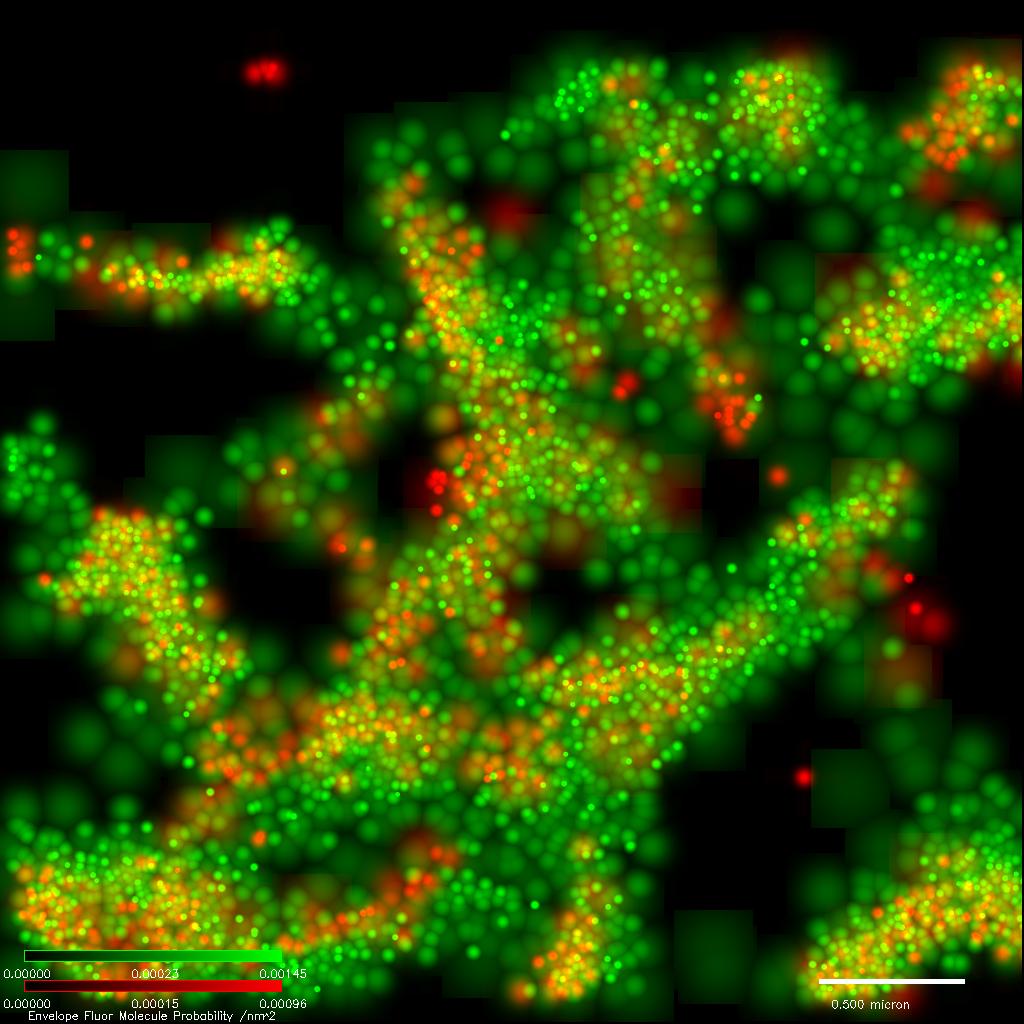


Sajman, J., Yakovian, O., Unger Deshet, N., Almog, S., Horn, G., Waks, T., Globerson Levin, A., Sherman, E. (2023). Nanoscale CAR organization at the immune synapse correlates with CAR-T effector functions. Cells 2023, 12(18), 2261
Wohl, I., Sajman, J. Sherman, E. (2023). Cell surface vibrations distinguish malignant from benign cells. Cells 2023, 12(14), 1901
Yakovian, O. Sajman, J., Alon, M., Arafeh, R., Samuels, Y., Sherman, E (2022). NRas activity is regulated by dynamic interactions with nanoscale signaling clusters at the plasma membrane. Iscience 25 (11), 105282
Wohl, I. Sherman, E. (2021). Reducing Myosin II and ATP-Dependent Mechanical Activity Increases Order and Stability of Intracellular Organelles. Int. J. Mol. Sci. 2021, 22(19), 10369
Sasson, E., , S., , B., , O., , M., , U., , B., , E., , G. D., Dzikowski, R., Ben-Zvi, A. (2021). Nano-scale Architecture of Blood-Brain Barrier Tight-Junctions. Elife 10, e63253.
Wohl, I. Sherman, E. (2021). Spectral Analysis of ATP-dependent mechanical vibrations in T cells. Front. Cell Dev. Biol. | doi: 10.3389/fcell.2021.590655
Sujman, J., Razvag, Y., Schidorsky, S., Kinrot, S., Hermon, K., Yakovian Y., and Sherman E. (2021) Adhering interacting cells to two opposing coverslips allows super-resolution imaging of cell-cell interfaces. Comm. Biology. 4, 439.
Yakovian, O., Sajman J., Arafeh, R., Neve-Oz, Y., Alon, M., Samuels, Y., Sherman, E. (2021). MEK Inhibition Reverses Aberrant Signaling in Melanoma Cells through Reorganization of NRas and BRAF in Self Nanoclusters. Cancer Research, 10.1158/0008-5472.CAN-20-1205.
Wohl, I., Yakovian, O. Sherman, E. (2020). Fluctuations in the Homogeneity of Cell Medium Distinguish Benign from Malignant Lymphocytes in a Cellular Model of Acute T Cells Leukemia. Appl. Sci. 2020, 10(24), 8894
Wohl, I., Sherman, E. (2020). Fast and synchronized fluctuations of cortical actin negatively correlate with nucleoli liquid-liquid phase separation in T cells. European Biophysics Journal 49, 409–423(2020)
Hermon, K., Time-correlated single molecule localization microscopy enhances resolution and fidelity. Scientific Reports, 10(1):16212.
Servili, E. Trus, M., Sajman, J., Sherman, E., Atlas, D. (2020). Elevated basal transcription can underlie timothy channel association with autism related disorders. Progress in Neurobiology. doi.org/10.1016/j.pneurobio.2020.101820
Razvag, Y., Neve-Oz, Y., Sajman, J., Yakovian, O., Reches M., Sherman, E (2019). T cell activation through isolated tight contacts. Cell Reports 29(11), P3506-3521.E6
Wohl, I., Sherman, E. (2019). ATP-Dependent Diffusion Entropy and Homogeneity in Living Cells. Entropy 2019, 21(10), 962
Razvag, Y., Neve-Oz, Y., Sherman, E., Reches M. (2018). Nanoscale Topography−Rigidity Correlation at the Surface of T Cells. ACSNano. DOI: 10.1021/acsnano.8b06366
Schidorsky, S., Yi, X., Razvag, Y., Sajman, J, Hermon, K., Weiss, S., Sherman, E. (2018). Synergizing superresolution optical fluctuation imaging with single molecule localization microscopy. Methods Appl. Fluoresc. 6, 045008
Neve-Oz, Y., Sajman, J., Razvag, Y., Sherman, E. (2018). InterCells: a generic Monte-Carlo simulation of intercellular interfaces captures nanoscale patterning at the immune synapse. Front. Immunol. 9:2051.
Razvag, Y., Neve-Oz, Y., Sajman, J., Reches, M., Sherman E. (2018). Nanoscale kinetic segregation of the T cell antigen receptor and CD45 in engaged microvilli facilitates early T cell activation. Nature Communications, 9:732.
Sajman, J., Trus, M., Atlas, D. 1, Sherman, E. 1 (2017). The L-Type Voltage-Gated Calcium Channel co-localizes with Syntaxin 1A in nano-clusters at the plasma membrane. Scientific Reports 7, 11350. 1-Corresponding authors
Golan, Y., Sherman, E. (2017). Resolving mixed mechanisms of protein subdiffusion at the T cell plasma membrane. Nature Communications, 8:15851.
Barr, V. A., Sherman, E., Yi, J., Akpan, I., Rouquette-Jazdanian, A. K., Samelson, L. E. (2016). Development of nanoscale structure in LAT-based signaling complexes. .
Schidorsky, S.*, Yi, X.*, Razvag, Y., Golan, Weiss, S.1, Sherman, E.1 Synergizing superresolution optical fluctuation imaging with single molecule localization microscopy. arXiv: 1603.04028. * - Equal contribution. 1 - Corresponding authors
Dubey, G. P.*, Mohan, G. B. M.*, Dubrovsky, A., Amen, T., Tsipshtein, S., Rouvinski, A., Rosenberg, A., Kaganovich, D., Sherman, E., Medalia, O., and Ben-Yehuda, S. (2016). Architecture and Characteristics of Bacterial Nanotubes. Developmental Cell, 36 (4) 452-61. * - Equal contribution.
Neve-Oz, Y. Razvag, J. Sajman, and E. Sherman. Mechanisms of localized activation of the T cell antigen receptor inside clusters. Biochim. Biophys. Acta. 1853 (2015), p. 810–21.
Parker J., Sherman E., van de Raa M., van der Meer D., Samelson L. E., & Losert W. (2013). Automatic sorting of point pattern sets using Minkowski Functionals. Phys. Rev. E., 88, 022720.
Sherman, E., Barr, V., Samelson, L. E. (2013). Resolving multi-molecular protein interactions by photoactivated localization microscopy. Methods, 59 (3), 261-9.
Sherman, E.*, Barr, V.*, Samelson, L. E. (2012). Super resolution imaging of signaling complexes downstream the T cell antigen receptor. Immunological Reviews. * - Equal contribution.
Sherman, E., Barr, V., Manley, S., Patterson, G., Balagopalan, L., Akpan, I., Regan, C.K., Merrill, R.K., Sommers, C.L., Lippincott-Schwartz, J., et al. (2011). Functional nanoscale organization of signaling molecules downstream of the T cell antigen receptor. Immunity 35, 705-720.
Sherman E. and Haran G. (2011). Fluorescence correlation spectroscopy of fast intra-molecular chain dynamics within denatured protein L. ChemPhysChem, 12(3) 696-703.
Balagopalan L.*, Sherman E.*, Barr V. A. and Samelson L. E. (2011). Imaging techniques for assaying lymphocyte activation in action. Nature Reviews Immunology 11, 21-33. * - Equal contribution.
Balagopalan L., Coussens N. P., Sherman E., Samelson L. E., and Sommers C. L. (2010). The LAT Story: A Tale of Cooperativity, Coordination, and Choreography, in Immunoreceptor Signaling. Cold Spring Harb Perspect Biol.
Phillip Y., Sherman E., Haran G. & Schreiber G. (2009). Common Crowding Agents Have Only a Small Effect on Protein-Protein Interactions. Biophysical Journal, Volume 97, Issue 3, 5, 875-885.
Aitziber C. L., Gregg L., Sherman E., Corey O. S., Regan L. & Haran G. (2008). Non-random coil behavior as a consequence of extensive PPII structure in the denatured state. Journal of Molecular Biology, Vol 382, Issue 1, 203-212.
Paz A., Zeev-Ben-Mordehai T., Lundqvist M., Sherman E., Mylonas E., Weiner L., Haran G., Svergun D. I., Mulder F. A. A., Sussman J. L. & Silman I. (2008). Biophysical Characterization of the Unstructured Cytoplasmic Domain of the Human Neuronal Adhesion Protein Neuroligin 3. Biophysical Journal, Vol 95, No. 4, 1928-1944.
Sherman E., Itkin A., Kuttner Y. Y., Rhoades E., Amir D., Haas E. & Haran G. (2008). Using Fluorescence Correlation Spectroscopy to Study Conformational Changes in Denatured Proteins. Biophysical journal, vol. 94, no12, 4819-4827.
Sherman E. & Haran G. Coil-globule transition in the denatured state of a small protein. (2006). Proc. Natl. Acad. Sci. USA 103(31) 11539-43.
Theme by Danetsoft and Danang Probo Sayekti inspired by Maksimer